Dr. Vibor Laketa

Infectious Diseases Imaging Platform (IDIP)
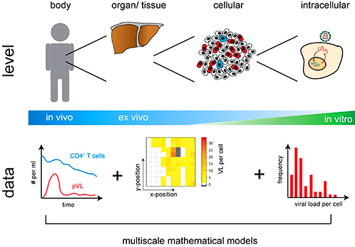
In CIID, we aim to achieve a comprehensive understanding of the physiology of major human pathogens and to develop improved therapeutic strategies through advanced microscopy-based molecular imaging, which can identify and quantitatively analyze individual molecular events underlying different pathophysiological states. Traditional biochemical, genetic, and “omics” approaches have provided valuable insights into host–pathogen interactions, but they are largely population-based, end-point analyses. Such methods often obscure rare or stochastic molecular events by averaging them out. Yet these individual events are precisely what drive the transition between healthy and diseased states. We believe that to truly understand different physiological states, we need to employ a reductionist experimental approach that is able to capture and quantitatively examine these individual molecular events. Such information can then be incorporated into mathematical models together with the observations made using the population-based approaches ultimately leading to a comprehensive understanding of the underlying patho-physiological processes.
Due to recent innovations, the only methodology able to capture and quantitatively examine individual molecular events in a complex biological system is microscopy-based imaging. A microscope is unique in being able to:
- show how “stuff” looks like (spatial organization of the living matter — molecules, organelles, cells, pathogens etc..)
- tell us how much “stuff” is where
- and how all this changes over time in a living system
A modern microscope is a robot that can sample complex dynamics of a biological system across a wide range of spatiotemporal scales and organizational levels of complexity, and thus is able to provide the most realistic representation of a living system. It is a big technical challenge to observe and quantify these stochastic molecular events. An equally big challenge is to understand how they give rise to different stereotypical patho-physiological states that we observe in “bulk” measurements. In this respect, microscopy-based analysis yields quantitative information that can be integrated into predictive multiscale mathematical models of infection that will ultimately be used to reconcile the findings obtained by the reductionist approach with the ones that are based on population measurements.

The Infectious Diseases Imaging Platform (IDIP) provides, develops and applies high-end microscopy infrastructure under enhanced biosafety containment 2 and 3 conditions (BSL‑2 and 3) to enable infectious diseases research.

Over 250m2 of ground (BSL‑2) and underground (BSL‑3) area of the CIID is dedicated to IDIP infrastructure which is organised in 14 interconnected microscopy rooms, tissue culture and sample preparation, image analysis and office areas. IDIP implements a comprehensive range of bioimaging technologies that allow infectious disease research across vastly different spatiotemporal scales and organizational complexities, going from structural studies on macromolecular scale all the way to whole organ/body imaging in living animals. More information about IDIP organisation and instrumentation can be found on the IDIP homepage https://www.idip-heidelberg.org/.

