

Dr. Vibor Laketa
Vibor.Laketa
@med.uni-heidelberg.de
Phone: +49 6221 56–34410
Establishment and Application of Advanced Microscopy Infrastructure
Establishment and Application of Advanced Microscopy Infrastructure
Projects
Microscopy and infectious disease research have been inseparable partners ever since in the 1670s Anthonie van Leeuwenhoek used his newly-invented microscope to examine a sample of plaque he had scraped from his own teeth, and observed — for the first time — bacteria and other microorganisms that share the world with us. Undoubtedly, microscopy was fundamental in infectious disease research, as it was necessary for the discovery of infectious agents by direct observation and later on also for their diagnosis.
In modern biomedical research that is being realized in CIID, microscopy-based experimental approach has a central role. A comprehensive understanding of host-pathogen interactions requires quantitative assessment of molecular events across a wide range of spatiotemporal scales and organizational complexities Due to recent technical developments, this is currently only achievable with microscopy. Based on development of new imaging modalities, fluorescent probes and sensors, computer technology, image analysis algorithms and new biological model system, the field of biological microscopy is undergoing a revolution. Only now we are starting to appreciate and make use of the full potential of the microscopy-based experimental approach. Modern microscopes have evolved into complex robotic instruments capable of automatically interrogating a wide range of biological processes at vastly different spatiotemporal scales and organizational complexities, from structural studies on macromolecular scale all the way to whole organ/body imaging in living animals.
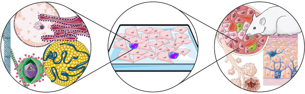
We believe that modern microscopy technology is currently uniquely positioned to propel the infectious disease research to a new frontier. For these reasons, we are establishing and applying advanced microscopy infrastructure to allow infectious disease research across the range of different spatiotemporal scales and organizational complexities.
More information about IDIP organisation and instrumentation can be found on the IDIP homepage https://www.idip-heidelberg.org/. If you would like to use the IDIP infrastructure, please follow the instructions at https://www.idip-heidelberg.org/usage-concept.
Microscopy Infrastructure under BSL2 and BSL3 Containment
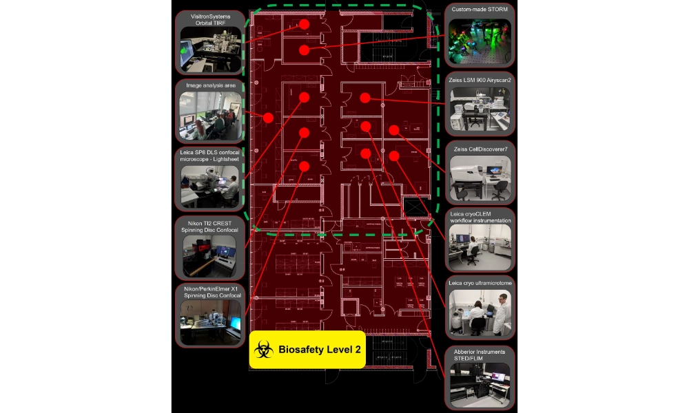
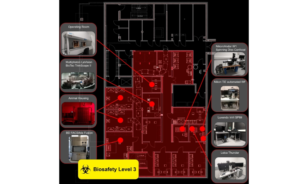
The Infectious Disease Imaging Platform (IDIP) is a scientific and technology platform that combines advanced microscopy and FACS infrastructure under enhanced biosafety conditions (BSL‑2 and BSL‑3) with project-specific expertise and targeted methodological development for infectious disease research, drug discovery and diagnostics. More than 250 m² of ground-level (BSL‑2) and underground (BSL‑3) space within the Center for Integrative Infectious Disease Research (CIID) is dedicated to IDIP, encompassing 14 interconnected microscopy rooms, tissue culture and sample preparation facilities, image analysis stations and office areas.
IDIP implements a comprehensive range of bioimaging technologies spanning vastly different spatial and temporal scales and levels of biological organization, from structural studies at the macromolecular scale to whole-organ and in vivo imaging. IDIP projects cover all aspects necessary for the execution of microscopy-based experimental approaches in infectious disease research. Activities typically include:
Implementing, operating and providing advanced microscopy and cell-sorting infrastructure under enhanced biosafety conditions (BSL‑2 and BSL‑3).
Consultation during microscopy-based project planning and experimental design.
Collaboration in microscopy-based projects conducted within CIID and beyond.
Advice and support in image processing, data visualization and presentation.
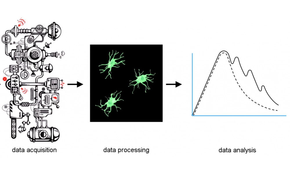
Development of automated data acquisition and image-analysis workflows.
$Coordination of microscopy infrastructure investments and support for related grant applications.
Education and training in microscopy and related technologies.
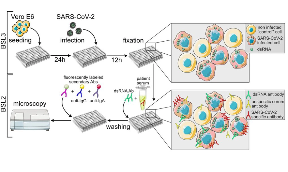
Development of microscopy-based assays for drug screening and diagnostics
Microscopy-based assays provide powerful approaches for advancing translational research, enabling high-content, single-cell analysis of complex biological systems with multiplexed, scalable and quantitative readouts. This creates new opportunities for drug discovery and for the development of sensitive, specific and adaptable diagnostic procedures, particularly in the context of emerging infectious diseases and therapeutic targets.
By combining advanced microscopy with automation, image analysis and machine learning, we develop microscopy-based assays capable of quantitatively characterizing complex cellular phenotypes and responses to candidate interventions. Through ongoing collaborations, these approaches are being applied to diverse biomedical challenges in infectious diseases and beyond, with the aim of developing robust, adaptable and physiologically relevant assay platforms for drug screening and diagnostics.
Heidelberg–Hamburg Infectious Diseases Imaging Association (HHH-IDIA)

HHH-IDIA is a DFG-funded collaborative initiative connecting the advanced imaging platforms in Heidelberg and Hamburg, two sites with a particularly broad and complementary portfolio of imaging technologies available under enhanced biosafety conditions. This combination provides a strong technological basis for advancing microscopy-based infectious disease research and addressing the specific challenges associated with imaging infectious material under BSL‑3 containment. The initiative brings together expertise in advanced microscopy, infection biology, biosafety and image analysis.
A central goal of HHH-IDIA is to improve the accessibility and application of advanced BSL‑3 imaging technologies within the infectious disease research community. Through collaboration between the participating sites, HHH-IDIA promotes methodological development, knowledge exchange and the harmonization of imaging approaches, strengthening the role of advanced microscopy in infectious disease research and pandemic preparedness. The Heidelberg site contributes advanced imaging expertise and infrastructure for the development and application of microscopy-based approaches in complex model systems. In close collaboration with industry partners, IDIP also evaluates the compatibility of advanced microscopy systems with decontamination procedures required for high-containment environments, helping to facilitate the safe implementation of emerging imaging technologies in BSL‑2 and BSL‑3 research.
Advanced Imaging and Cell Sorting for Synthetic Immunology (SynthImmune)
SynthImmune is a Cluster of Excellence at Heidelberg University dedicated to developing the emerging field of bottom-up synthetic immunology. The initiative combines approaches from synthetic biology, nanotechnology, systems biology and immunology to understand, engineer and reconstitute immune functions, with the long-term goal of developing new strategies for the prevention and treatment of infectious diseases and cancer.
Within SynthImmune, IDIP serves as a technology platform for advanced microscopy and fluorescence-activated cell sorting under enhanced biosafety conditions (BSL‑2 and BSL‑3). The platform combines specialized imaging and cell-sorting technologies with support throughout the experimental workflow, from experimental design and method development to data acquisition, quantitative image analysis and interpretation.
A particular focus is the development and application of imaging-based approaches to identify highly functional immune cells and characterize their behaviour in physiologically relevant model systems. Advanced microscopy enables quantitative analysis of immune-cell morphology, dynamics and interactions, including in complex three-dimensional and microphysiological models. In combination with automated image analysis and cell sorting, these approaches support the identification and functional selection of rare immune-cell populations and provide new opportunities for linking cellular phenotypes to immune function.
Advanced Imaging of Viral Infection and Antiviral Drug Discovery (SHIELD)

SHIELD – Molecular Strategies against Viral Entry and Glycan Shielding is a Horizon Europe research project investigating mechanisms of viral entry, virion glycosylation and immune evasion, with the aim of identifying and validating strategies to interfere with viral infection. By combining molecular modelling, structural biology, virology, cellular systems and advanced imaging, the consortium investigates viruses with epidemic and pandemic potential, including Lassa virus (LASV), Hendra virus (HeV), Nipah virus (NiV), and the flaviviruses dengue (DENV), West Nile (WNV), Zika (ZIKV) and yellow fever (YFV).
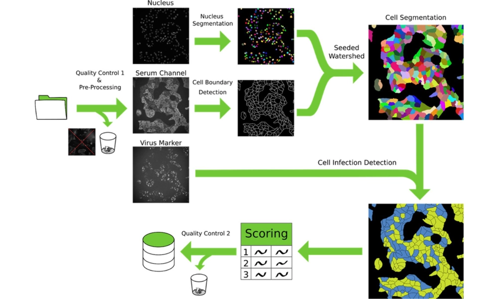
Within SHIELD, IDIP develops and applies advanced imaging approaches to investigate viral infection dynamics across different stages of the viral life cycle, including virus–cell interactions, entry, intracellular processes and the spread of infection in cellular and complex model systems. A major component of the IDIP contribution is the development of automated and AI-assisted workflows for image acquisition and analysis, enabling unbiased quantitative measurements and increased assay throughput. IDIP further contributes to the benchmarking and validation of assay methodologies across the SHIELD consortium through the exchange of experimental materials, inter-laboratory comparisons and systematic assessment of assay performance. These activities support the establishment of robust and transferable experimental pipelines for the evaluation of antiviral compounds.
Advanced Imaging for Translational Infectious Disease Research (DZIF)
The German Center for Infection Research (DZIF) brings together basic and clinical research with the goal of translating scientific discoveries into improved strategies for the prevention, diagnosis and treatment of infectious diseases. A central challenge in this process is to bridge the gap between mechanistic studies in simplified experimental systems and the complex biological processes occurring in physiologically relevant tissues and organisms.
Within DZIF, IDIP supports research in the TTU HIV and other DZIF Thematic Translational Units, combining advanced imaging technologies with experimental consultation, methodological development and quantitative image analysis. The platform enables quantitative investigation of pathogen behaviour and host–pathogen interactions in complex, close-to-physiological experimental systems, generating spatially and temporally resolved data with increased translational potential.
By enabling infectious disease research in increasingly physiologically relevant model systems, IDIP contributes to bridging the gap between mechanistic studies and translational research. Such approaches can provide a more direct link between preclinical findings and clinically relevant biology, while also creating a technological foundation for rapidly investigating emerging pathogens and developing new approaches for infectious disease preparedness and response.