


Dr. Marina Lusic
Group leader and W2 tenure-track DZIF Professor for preclinical HIV‑1 research
Integrative Virology, Center for Integrative Infectious Disease Research (CIID), Medical Faculty Heidelberg, Heidelberg University
marina.lusic@med.uni-heidelberg.de
ORCID 0000–0002-0120–3569
Nuclear architecture in viral infection
Nuclear architecture in viral infection
Research
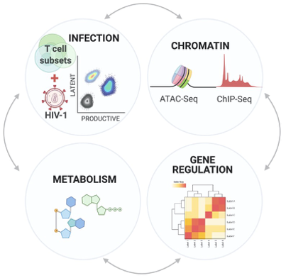
Our lab explores how nuclear architecture and cellular metabolism respond to HIV‑1 infection. The nucleus is a highly organized environment where RNA and chromatin structure support essential cellular functions. When HIV‑1 enters a cell, its genome must integrate into cellular DNA to establish a productive infection, and the site of integration influences viral transcription and replication — shaping the course of infection.
At the same time, viral replication induces cellular stress at both the metabolic and chromatin levels. The interplay between these factors determines whether the virus keeps replicating or becomes dormant, persisting in the host genome despite antiviral treatment. To study these processes we use primary models of T cells and are establishing cultures of primary human microglia, applying state-of-the-art genomics and microscopy to follow HIV‑1 integration and latency.
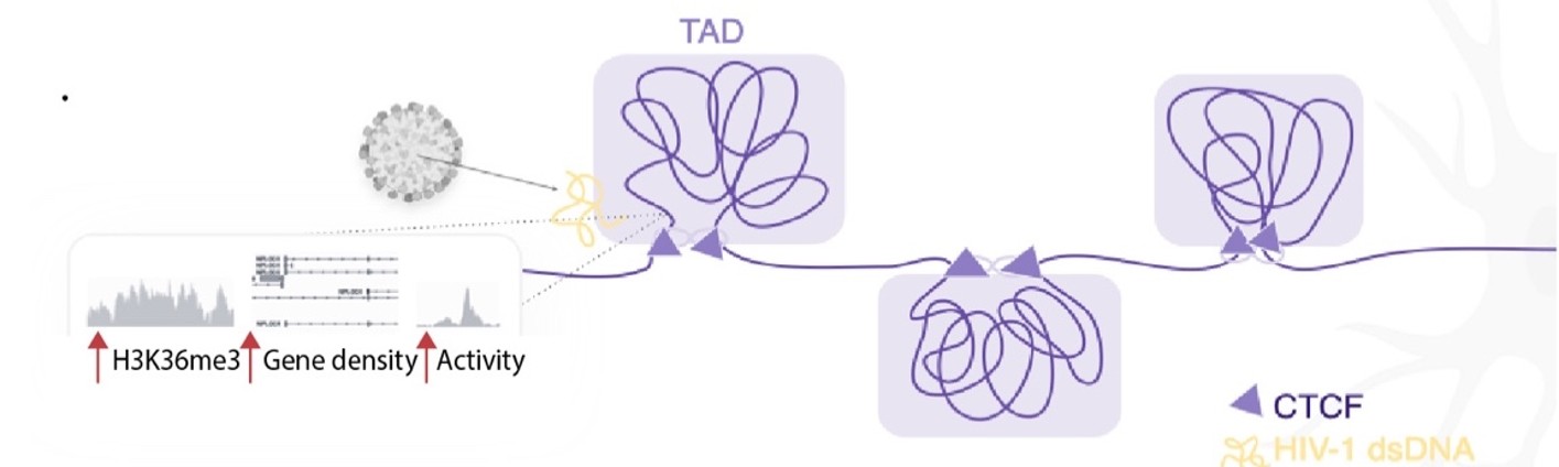
1 | Genome organization and chromatin in HIV‑1 integration
Sites of HIV‑1 integration are essential determinants of viral fate. Where in the cellular genome HIV‑1 integrates will define whether and how will the virus be silenced and whether or not could it be reactivated. We are interested in understanding where HIV‑1 inserts into the host genome, and how those integration sites are arranged in three-dimensional nuclear space — from recurrent target genes and super-enhancer clusters to CTCT and TAD boundaries (Marini et al Nature 2015, Lucic et a Nat Commun. 2019, Michieletto et al Nat Commun. 2019, Rheinberger et al. Cell Reports 2023). Using 3D immuno-DNA FISH together with genome-wide mapping, we follow these rules in primary CD4+ T cells and in microglia, the long-lived reservoir of the central nervous system We combine our genomic and bioinformatic analysis with physical modelling (in collaboration with Andrea Maria Chiariello and Andrea Fontana) to understand how HIV‑1 selects it’s preferred chromatin environment.
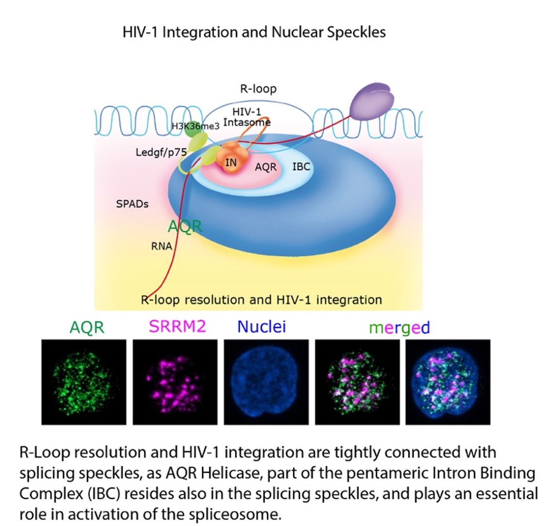
2 | RNA:DNA hybrids and splicing
Our recent findings show that HIV‑1 prefers to integrate at genomic features rich in RNA:DNA hybrids (R‑loops) and near nuclear speckles, the compartments where splicing is organized. We showed that the splicing helicase Aquarius, part of the Intron Binding Complex, resolves these R‑loops at sites of integration in speckle-associated, H3K36me3-marked chromatin engaged by LEDGF/p75 (Penzo et al. Nat Microb 2025; Müller et al. EMBO J 2025). These events link where the virus lands to how host and viral genes are spliced and expressed. We are particularly interested in how these genomic features and splicing factors, also known also to be involved in DNA repair might contribute to the intactness of the proviral genome. At the same time, we are exploring the relevance of the newly described involvement of the RNA portion of chromatin (R‑loops) for the function of HIV‑1 integrase allosteric inhibitors (Penzo et al, in preparation).
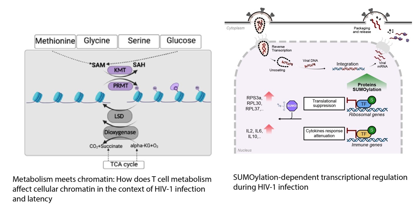
3 | T‑cell metabolism, chromatin and latency
The main obstacle to a functional HIV cure is the latent reservoir of provirus established in memory CD4+ T cells, which cannot be eradicated with current antiretroviral treatment. Our work was the first to point to PML Nuclear Bodies as the nuclear architectural shelter of latent HIV‑1 genomes (Lusic et al. Cell Host Microbe 2013, Lucic et al. Adv Science 2025). While dissecting the molecular mechanisms that establish and maintain latency, we found that the oxidative phosphorylation pathway and cellular oxidative stress play a central role in HIV‑1 transcriptional control, coupled with the disruption of PML Nuclear bodies during acute infection, and their reformation during latency establishment (Shytaj, Lucic et al. EMBO J 2020). Concomitantly, during latency establishment infected cells also downregulate the glycolytic pathway (Shytaj et al. EMBO Mol Med 2021), pointing to the very intriguing interplay between cellular metabolism and chromatin organization. Two projects are following these findings- in one, we characterize cellular metabolites and find a dysregulation of the methionine pathway with consequences for chromatin and HIV‑1 reactivation (Giardina and Lucic, in preparation); in the other, we describe SUMOylation-dependent transcriptional regulation in HIV‑1 infected CD4+ T Cell (Giardina and Lucic, in preparation).
4 | Genome organization at the borders
A new strand of the lab’s work asks whether the nuclear periphery is a single environment or several. We find that active, super-enhancer–marked genes — including HIV-1’s own target genes — shift toward the periphery when CD4+ T cells are activated, apparently toward nuclear pores rather than the repressive lamina.
By mapping the genome associated with the basket nucleoporin Nup153 and following individual gene alleles as cells activate (de Castro et al. BioRXIV 2024), we are testing whether a permissive, pore-organized compartment sits alongside the repressive lamina — and whether pore number itself helps set how the genome is arranged.
These questions are part of a broader move toward the chromatin biology of primary human T cells at molecular resolution. Working with the Beck and Hummer laboratories, we recently used in-situ cryo-electron tomography to resolve individual nucleosomes and map the architecture of heterochromatin at the nuclear periphery of resting T cells — a step toward reading genome organization directly, structure by structure (Kreysing et al., Nature Communs 2026).
Selected Publications
Complete publication list (PubMed)
2026 Kreysing JP, Cruz-León S, Betz J, et al., Lusic M, Hummer G, Beck M. Molecular architecture of heterochromatin at the nuclear periphery of primary human cells. Nature Communications 17.
2025 Penzo C, Ozel I, Kuzman M, et al., Parissi V, Lusic M. Aquarius helicase facilitates HIV‑1 integration into R‑loop–enriched genomic regions. Nature Microbiology.
2024 de Castro IJ, Schuster L, Patino Gomes C, et al., Saka S, Lusic M. Nucleopore complex harbors splicing machinery for efficient mRNA processing. bioRxiv.
2023 Rheinberger M, Costa AL, Kampmann M, et al., Herrmann C, Lusic M. Genomic profiling of HIV‑1 integration in microglia links viral integration to topologically associated domains. Cell Reports 42:112110.
2022 Shytaj IL, Feres M, Lucic B, Galluci L, et al., Lusic M. The The FDA-approved drug cobicistat synergizes with remdesivir to inhibit SARS-CoV‑2 replication in vitro and decreases viral titers and disease progression in Syrian hamsters. mBio 2022 Apr 26:13(2).
2021 Zila V, Margiotta E, Turoňová B, et al., Lusic M, Kräusslich HG, Beck M. Cone-shaped HIV‑1 capsids are transported through intact nuclear pores. Cell 184.
2019 Lucic B, Chen H‑C, Kuzman M, et al., Filion GJ, Lusic M. Spatially clustered loci with multiple enhancers are frequent targets of HIV‑1. Nature Communications 10:4059.
2017 Lusic M, Siliciano RF. Nuclear landscape of HIV‑1 infection and integration. Nature Reviews Microbiology 15:69–82.
2015 Marini B, Kertész-Farkas A, Ali H, et al., Giacca M, Lusic M. Nuclear architecture dictates HIV‑1 integration site selection. Nature 521:227–231.
2013 Lusic M, Marini B, Ali H, Lucic B, Luzzati R, Giacca M. Proximity to PML nuclear bodies negatively regulates HIV‑1 gene expression in CD4+ T cells. Cell Host & Microbe 13:665–677.